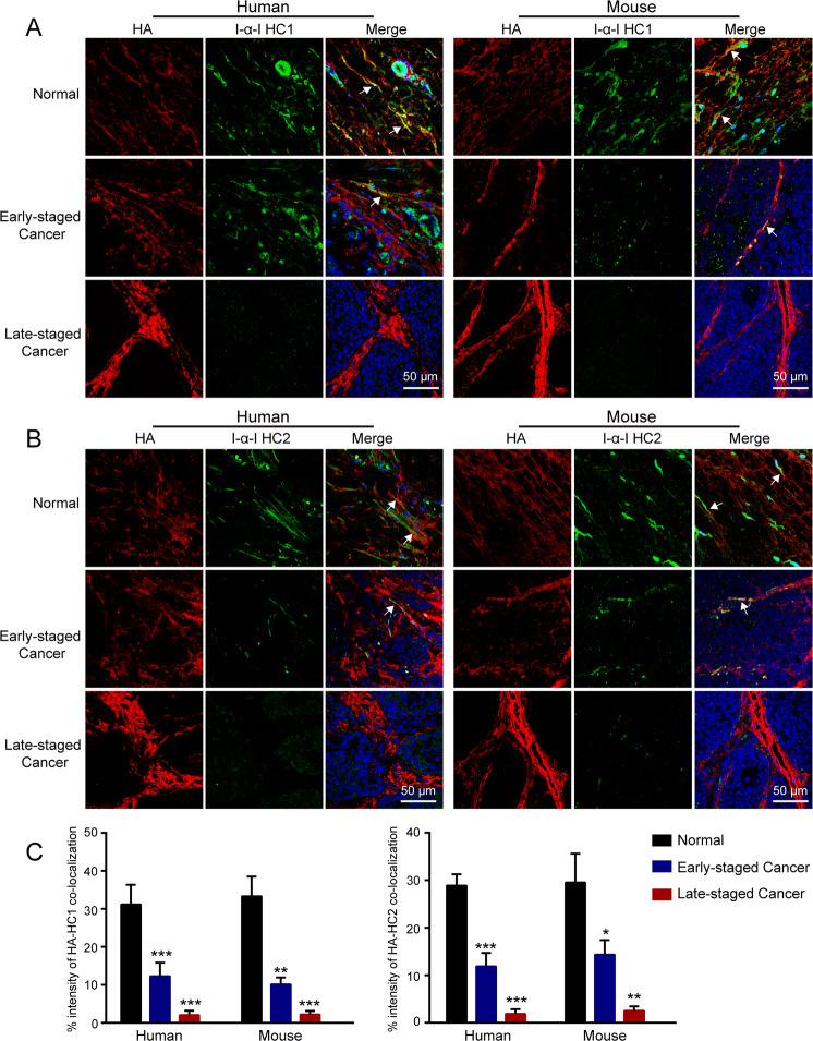
Fig. 1. HA cross-linking was decreased in breast cancer tissues and associated with tumor stage.
A Immunofluorescence staining of HA (red) and I-α-I HC1 (green) in cancer tissues and corresponding adjacent normal tissues from patients with early (stage I, n = 7) or late (stage III, n = 9) staged breast cancer, normal mammary gland tissues from FVB mice, and cancer tissues from MMTV-PyMT mice with early (week 10) or late (week 16) staged breast cancer. B Immunofluorescence staining of HA (red) and I-α-I HC2 (green) in corresponding tissues. The white arrowheads refer to the co-localization of HA and I-α-I, which means HA cross-linking. HA and I-α-I HCs were both localized within and outside of cells. C The intensity ratio of HA co-localized with I-α-I HCs to total HA was used to determine the levels of HA-HC complexes. The intensity percentage of HA-HCs co-localization was acquired by the Image J software. Error bars show mean ± SD values. Statistical analysis was performed using Student’s t test. *p < 0.05, **p < 0.01, ***p < 0.001.

