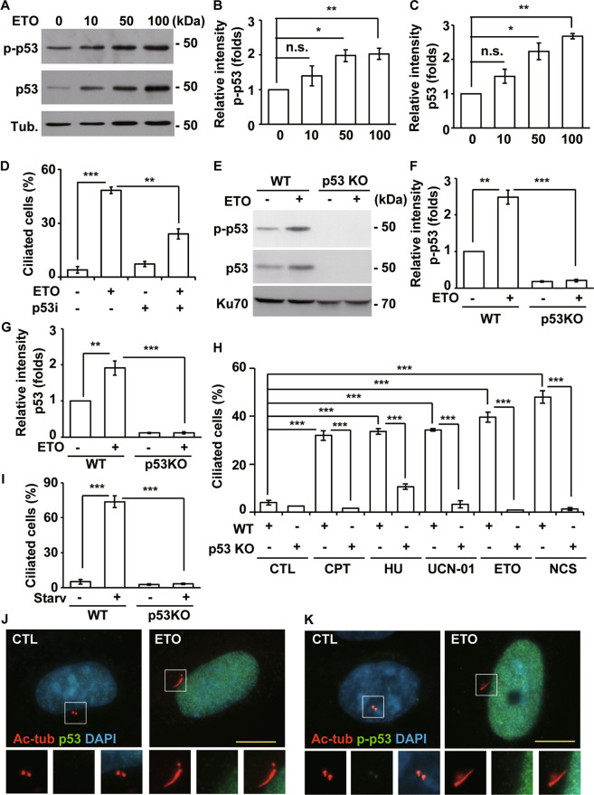
Fig. 4. ETO induces p53 activation to promote ciliogenesis in RPE1 cells.
A ETO activated p53 in RPE1 cells. Extracts of cells treated with different concentration of ETO for 24 h were analyzed with antibodies against phosphorylated p53 (p-p53), p53, and tubulin (Tub.). Quantitative results of relative intensity of p-p53/actin (B) and p53/actin (C) of A. All ETO-treated data were normalized to the data without ETO treatment. D Inhibition of p53 reduced ETO-induced ciliogenesis. Quantitative results of frequency of ciliated RPE1 cells treated with 100 µM ETO for 24 h in the presence or absence of p53 inhibitor, pifithrin-α. ETO-induced ciliogenesis were inhibited in p53 knockout RPE1 cells. E Extracts of wild-type (WT) or p53 knockout (p53 KO) cells treated with ETO for 24 h were analyzed with antibodies against p53 and Ku70. Quantitative results of relative intensity of p-p53/actin (F) and p53/actin (G) of E. H Knockout of p53 inhibited ciliogenesis. Quantitative results of frequency of ciliated RPE1 cells treated with cisplatin (CPT), hydroxyurea (HU), 7-Hydroxystaurosporine (UCN-01), ETO, and neocarzinostatin (NCS) for 24 h. I Knockout of p53 inhibited starvation (Starv)-induced ciliogenesis. Quantitative results of the frequency of ciliated RPE1 cells under serum starvation for 24 h in wild-type and p53 knockout cells. Total p53 (J) and phosphorylated p53 (K) did not localize to the cilia and centriole in ETO-treated RPE1 cells. Immunostaining of CTL- or ETO-treated RPE1 cells with antibodies against p53 (J) or p-p53 (K) and acetylated tubulin (Ac-tub). DNA was stained with DAPI (blue). Scale bar, 10 µm. *P < 0.05; **P < 0.01; ***P < 0.001; n.s. no significance.

