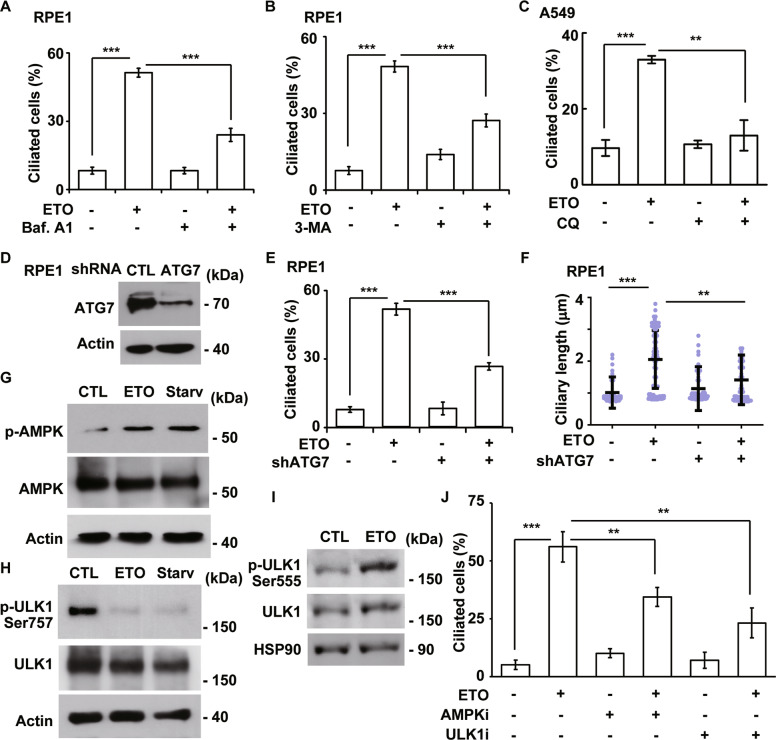
Fig. 6. ETO-activated autophagy promotes ciliogenesis.
ETO-induced autophagy facilitates ciliogenesis. Inhibition of lysosomal degradation by Baf.A1 (A) or autophagy initiation by 3-MA (B) reduced the frequency of ciliated RPE1 cells upon ETO treatment. C Inhibition of lysosomal degradation by CQ inhibited the frequency of ciliated A549 cells upon ETO treatment. Depletion of ATG7 by transfection of lentivirus containing shRNA against ATG7 (shATG7) inhibited ciliogenesis upon ETO treatment. D ATG7 was efficiently depleted. Extracts of RPE1 cells infected with shATG7 lentivirus were analyzed by immunoblotting with antibodies against ATG7 and actin. Quantitative results of the frequency (E) and length (F) of ciliated control and ATG7-deficient RPE1 cells in the absence or presence of ETO. G–I ETO treatment activated AMPK-ULK1 complex. Extracts of RPE1 cells treated with control (CTL), ETO, or serum starvation (Starv) for 24 h were analyzed with antibodies against phosphorylated AMPK (at Thr172), AMPK, phosphorylated ULK1 (at Ser757 or Ser555), actin, and HSP90. J Inactivation of AMPK and ULK1 reduced ETO-induced ciliogenesis. Quantitative results of the frequency of ciliated control, dorsomorphin- (5 μM; AMPKi), or SBI-0206965- (10 μM; ULK1i) treated RPE1 cells in the absence or presence of ETO. The results are presented as the mean ± SD of three independent experiments; more than 100 cells were counted in each individual group. **P < 0.01; ***P < 0.001.

