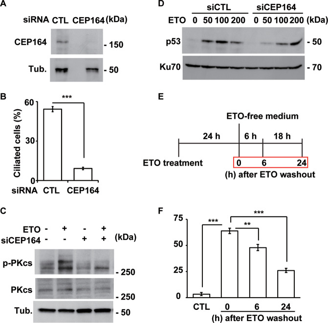
Fig. 8. The primary cilium maintains the ETO-induced DNA damage response.
Inhibition of ciliogenesis decreased ETO-induced DNA-PK activation. A Depletion of CEP164 inhibited ETO-induced DNA-PK activation. Extracts of RPE1 cells transfected with siRNA against CEP164 in the presence of ETO were analyzed by immunoblotting with antibodies against CEP164 and tubulin (Tub.). B Quantitative results of the frequency of ciliated CEP164-deficient RPE1 cells in the presence of ETO. The results are presented as the mean ± SD of three independent experiments; more than 100 cells were counted in each individual group. C Extracts of RPE1 cells transfected with siRNA against CEP164 were analyzed by immunoblotting with antibodies against phosphorylated DNA-PKcs (p-PKcs), DNA-PKcs (PKcs), and tubulin (Tub.). D Depletion of CEP164 inhibited ETO-induced p53 activation. Extracts of RPE1 cells transfected with siRNA against CEP164 in the presence of different concentrations of ETO were analyzed by immunoblotting with antibodies against p53 and Ku70. Removal of ETO reduced ETO-induced ciliogenesis in a time dependent manner. E Schematic representation of the experimental procedure used for F. Cells were treated with ETO for 24 h followed by culturing in ETO-free medium for additional 6 or 24 h before harvesting. F Quantitative results of the frequency of ETO-treated ciliated RPE1 cells after ETO washout. The results are presented as the mean ± SD of three independent experiments; more than 100 cells were counted in each individual group. **P < 0.01; ***P < 0.001.

