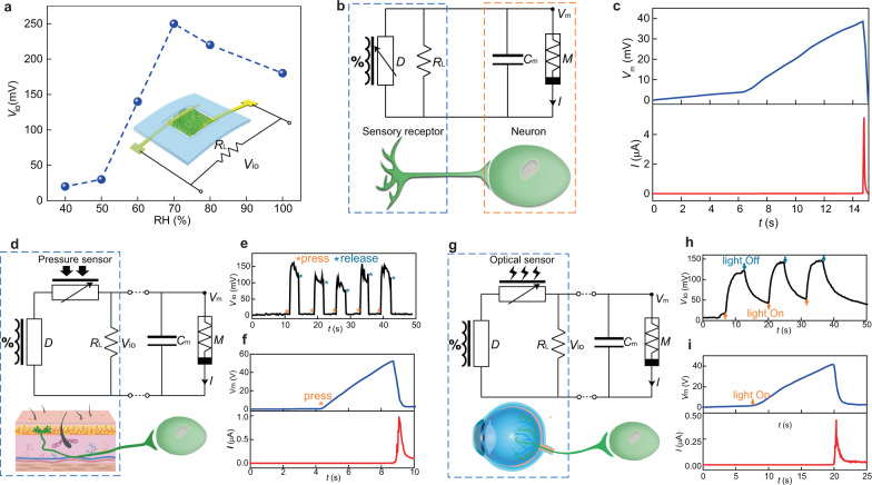
Fig. 2. Multifunctional sensory neuromorphic interfaces.
a Voltage output (Vio) from a vertical protein nanowire sensor connected with a load resistor (RL) measured at different RH. b Circuit diagram of feeding Vio from the protein nanowire sensor (D) into an artificial neuron made from a protein nanowire memristor (M) and a capacitor (Cm) by emulating an afferent circuit (bottom). c Evolution in the membrane potential (Vm) and current (I) in the artificial neuron when placed the sensor on a sweating skin. d Circuit diagram of feeding Vio from a tactile sensory component (made from a protein nanowire device (D) and a resistive pressure sensor) into an artificial neuron by emulating a tactile afferent circuit (bottom). e Touch-induced Vio from the tactile sensory component. f Evolution in the membrane potential (Vm) and current (I) in the artificial neuron by pressing the pressure sensor. g Circuit diagram of feeding Vio from an optical sensory component (made from a protein nanowire device (D) and an optical sensor) into an artificial neuron by emulating an optical afferent circuit (bottom). h Light-induced Vio from the optical sensory component. i Evolution in the membrane potential (Vm) and current (I) in the artificial neuron by lighting the optical sensor.

