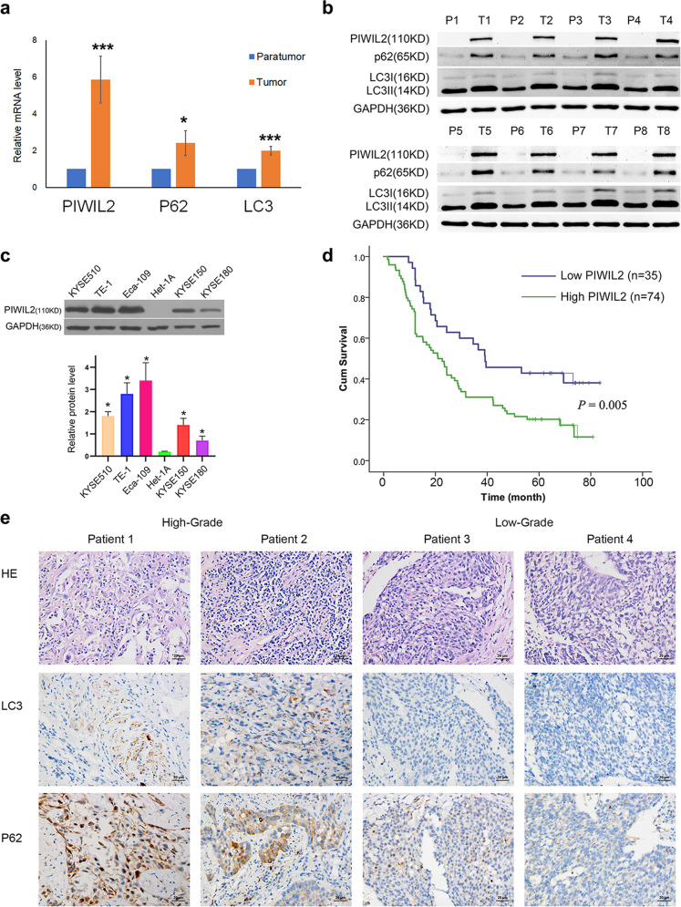
Fig. 1. PIWIL2 is highly expressed and associated with poor prognosis in ESCC.
Total mRNAs and proteins were extracted from tissue samples of eight individual ESCC patients, and subjected to RT-PCR (a) and western blot (b) respectively. T, tumor tissues; P, paratumor tissues. c ESCC cell lines (KYSE150, KYSE180, KYSE510, and Eca-109) and normal esophageal epithelial cell line Het-1A were subjected to expression analysis of PIWIL2 using western blotting (WB). d Kaplan–Meier curve depicting the long-term survival of the ESCC patients (n = 109). The curves were stratified based on the PIWIL2 level scored by intensity (0–3) and area (0–4) of the staining with TMA and IH technology (Log-rank test, p = 0.005). e Patients were randomly selected from PIWIL2 high-expression group and low-expression group (two from each). Biomarkers of autophagy (LC3 and P62) were analyzed using immunohistochemistry. Scale bar, 20 µm. *p < 0.05; ***p < 0.001.

