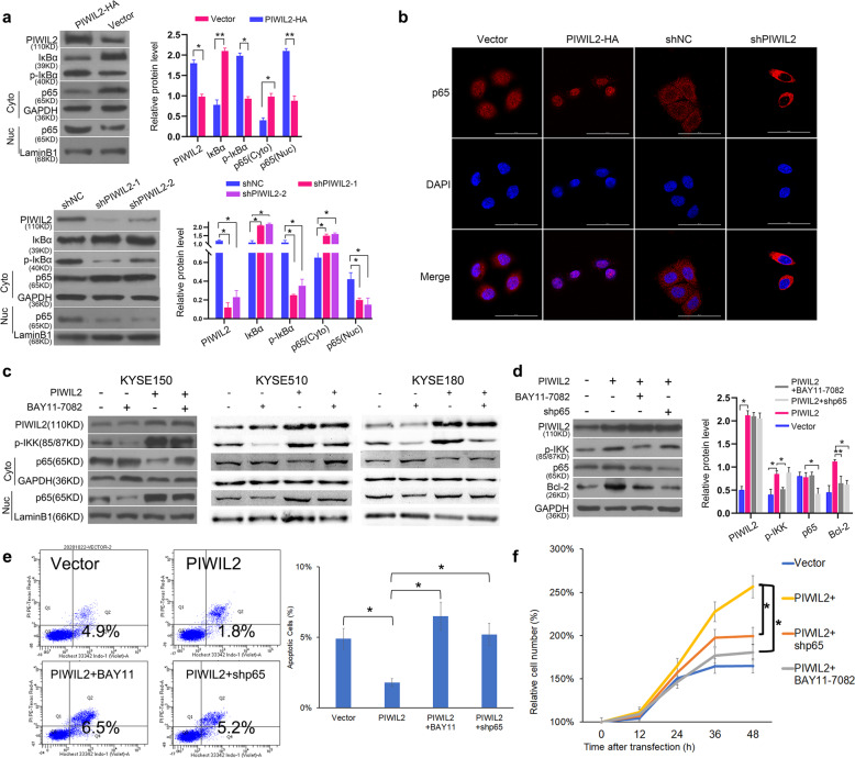
Fig. 4. PIWIL2 activates NF-κB signaling pathway through IKKβ to regulate apoptosis.
a KYSE150 cells were harvested 48 h after transfection and fractionated into nuclear (Nuc) and cytoplasmic (Cyto) components. The translocation of NF-κB were analyzed by measuring p65 subunit level in each component using WB. GAPDH and LaminB1 were employed as internal control, respectively. b Immunofluorescence microscopy for localization of p65 in KYSE150 cells 48 h after transfection. The nuclei were stained with 1 μg/ml DAPI (blue). Scale bar, 50 µm. c Transfected cells were treated with 10 μM IKK inhibitor BAY11-7082 for 12 h before nuclear/cytoplasmic fractionation. d KYSE150 cells with IKK knockdown were treated with BAY11-7082. Autophagic markers (LC3-II and p62) were examined to evaluate the selectively inhibition of BAY11-7082 on IKK. e Starvation induced apoptosis was analyzed by flow cytometry with Hochest33342/PI double staining. Apoptosis(%) = percentage of Hochest+ve/PI−ve cells. f Proliferation rates of transient transfected KYSE150 cells were revealed by crystal violet assay. Mock-transfected cells were used as control. *p < 0.05; **p < 0.01.

