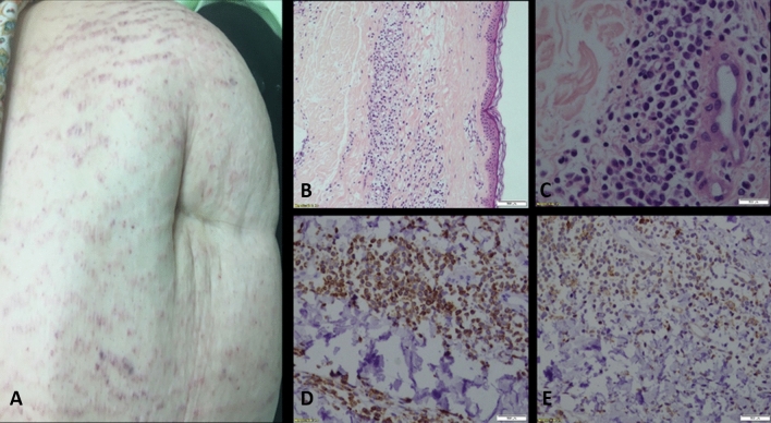
Figure 1.
Leukemia cutis. A 42-year-old woman with AML who underwent biopsy of a skin eruption. (A) The patient presented with hyperleukocytosis and abdominal purple striae-like infiltrated plaques in a reticulated pattern. (B,C) A punch biopsy performed from the right abdomen revealed a mild perivascular infiltrate in the dermis, composed of medium-sized immature atypical cells, otherwise unremarkable. The epidermis shows no significant changes; note the subepidermal Grenz zone (H&E, X100). (C) Same as (B) at higher magnification (H&E X400). (D,E) The atypical cells stain positive for CD45 (X200) (D) ,CD4 (X200)) E) ,HLA-DR, CD11c, CD68 and lysozyme. Cells are negative for CD3, CD20, CD79a, CD34, CD138, TdT, CD30, CD123 and cytokeratin. Ki67 shows a proliferative index of 25% in the infiltrates. The overall histological findings together with the immunohistochemical stains are compatible with AML involving the skin (leukemia cutis).

