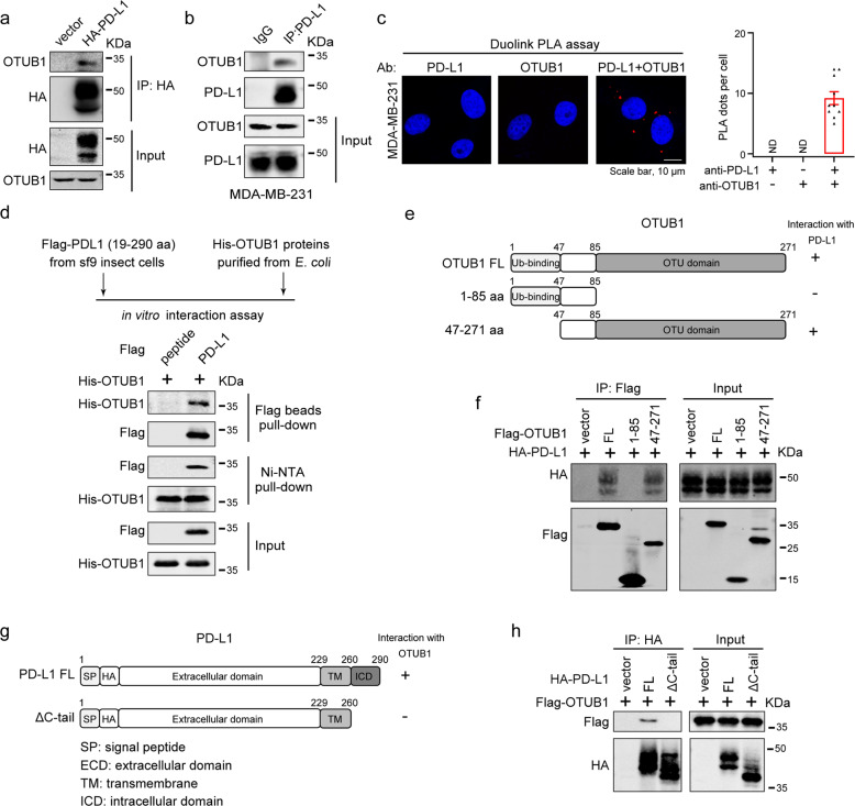
Fig. 2. OTUB1 specifically interacts with PD-L1 in vivo and in vitro.
a HEK293T cells were transfected with HA-PD-L1 or HA vector, and immunoprecipitation was performed with anti-HA antibody to examine the interaction between HA-PD-L1 and endogenous OTUB1. b Association between endogenous OTUB1 and PD-L1 in MDA-MB-231 cell lines was detected by Co-IP assays using IgG or PD-L1 antibodies. c In situ interaction between OTUB1 and PD-L1. Cells were fixed with 4% paraformaldehyde, immunostained with OTUB1 and PD-L1 antibodies and then assessed using the Duolink PLA assay. Scale bar, 10 µm. Quantification of the PLA dots indicating PD-L1-OTUB1 interactions was shown as mean ± SEM. d The direct interaction between recombinant Flag-PD-L1 19-290 aa proteins and His-OTUB1 proteins examined by in vitro pull-down assays. e Schematic representation of various OTUB1 truncations. f Mapping of OTUB1 domains critical for PD-L1 binding. HEK293T cells were transfected with different OTUB1 truncations, and cell lysates were immunoprecipitated with anti-Flag antibody to detect their PD-L1 binding ability. g Schematic representation of PD-L1 full-length and ΔC-tail constructs. h The interaction between Flag-OTUB1 and PD-L1 full-length or ΔC-tail detected by Co-IP assays.

