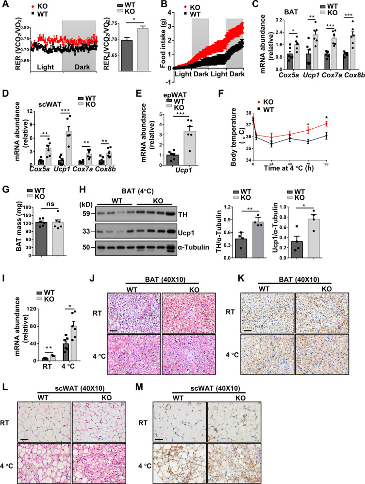
Fig. 6. Macrophages deficient in Kdm2a increase energy expenditure and adaptive thermogenesis.
A Results for real-time recording of RER (left) and quantitative data (right) collected from four mice in each group for 24 h following 16 weeks of HFD induction (n = 4 per group). B Food intake (g) in WT and KO mice following 16 weeks of HFD induction measured in metabolic cages (n = 4 per group). RT-PCR results for analysis of thermogenic genes Cox5a, Ucp1, Cox7a, and Cox8b in BAT (C) and scWAT (D) from HFD challenged WT and KO mice (n = 6 for each group). E RT-PCR results for analysis of Ucp1 in the epWAT following 16 weeks of HFD challenge (n = 6 for each group). F Daily rectal temperature of mice housed under 4 °C condition (n = 6 per group). G Analysis of the weight for BAT collected from WT and KO mice after cold exposure (n = 6 per group). H Representative western blot results (left) and bar graphs (right) showing the expression levels for BAT TH and Ucp1 (n = 4 per group). I Relative mRNA abundance of Ucp1 in BAT of mice with (n = 6/group) or without cold stress (n = 3/group). Representative images of H&E staining (J) and Ucp1 IHC (K) of BAT sections with or without cold exposure (six images per mouse). Representative images of H&E staining (L) and Ucp1 IHC (M) of scWAT sections (six images per mouse). Scale bars: 50 μm (J–M). Original magnification: ×400 (J–M). Values are expressed as mean ± SEM, and unpaired Student’s t test was used for data analysis. *P < 0.05; **P < 0.01; ***P < 0.001. ns not significant.

