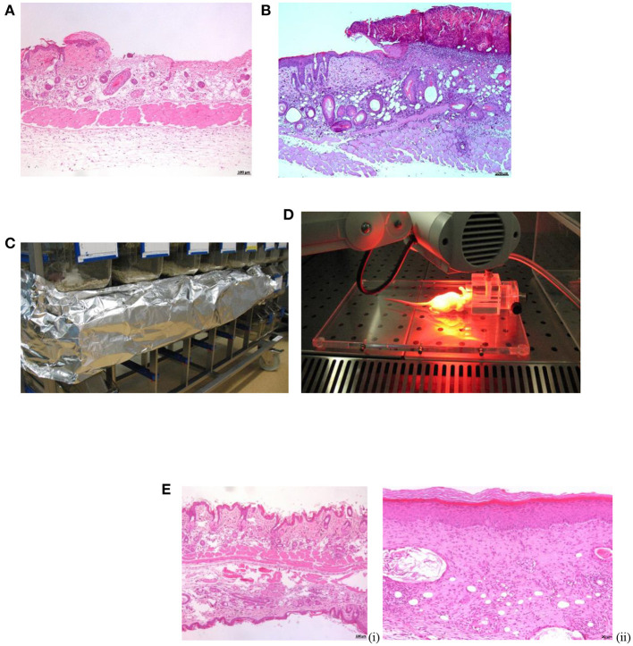
Figure 1.
Procedures in SKH-1 mice. (A) Microphotograph of skin abrasion experimentally induced. Note the very superficial loss of the epidermis. H-E. x50. (B) Microphotograph of the purulent crust observed at 24–48 h post-infection. H-E. x50. (C) Incubation of BM (aPDT) on dark. (D) MBa-PDT session. (E) Microphotograph of SKH-1 mice healthy skin (i) vs. skin healed per se (S. aureus infection) at 13 days post-inoculation (ii). H-E. x50.

