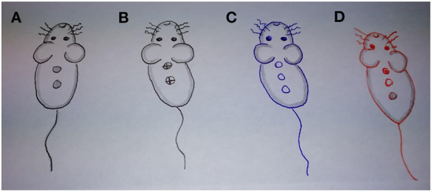
Figure 2.

Sampling of murine wounds (handmade) for comparisons. (A). Infected wounds treated with MB-aPDT. (B) Infected wounds treated with MU (dorsally combined with aPDT and ventrally in solitary). (C) Control (untreated) Infected wounds. (D) Healthy skin (dorsally ulcer by abrasion, in the middle intact skin, ventrally intact skin with MB).
