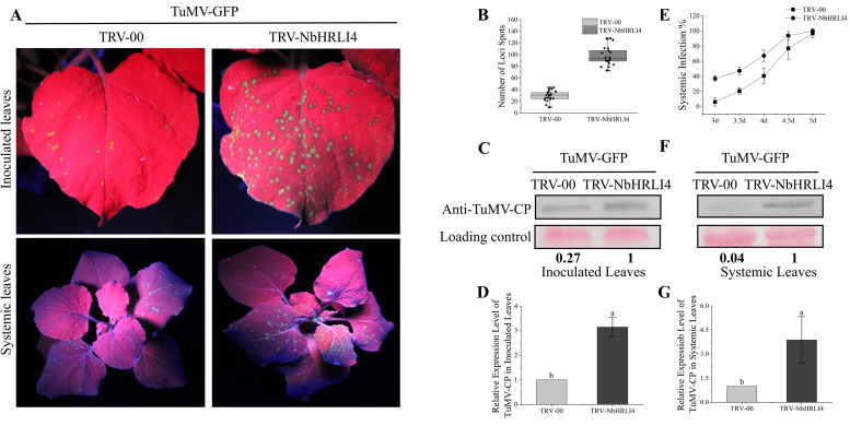
FIGURE 3.
Silencing of NbHRLI4 induced TuMV accumulation. (A) Inoculated and systemic leaves of plants inoculated with TuMV-GFP are observed under UV light. (B) Number of infection spots after TuMV infection of leaves inoculated with TRV-00 or TRV-HRLI4 (at least 20 plants per replicate) at 4 dpi. (C,D) Western blotting and RT-qPCR showing the increased expression of TuMV-CP (C) and mRNA (D) in inoculated leaves from TRV-NbHRLI4-infected plants compared with the TRV-00 infected plants at 3 dpi. (E) Percentage of plants systemically infected at different times after TuMV inoculation (at least 10 plants per replicate). (F,G) Western blotting and RT-qPCR showing the increased expression of TuMV-CP (F) and mRNA (G) in systemic leaves from TRV-NbHRLI4-infected plants compared with the TRV-00-infected plants at 5 dpi. The mean expression values in this figure were analyzed using t-test. Different letters on histograms indicate significant differences (P < 0.05).

