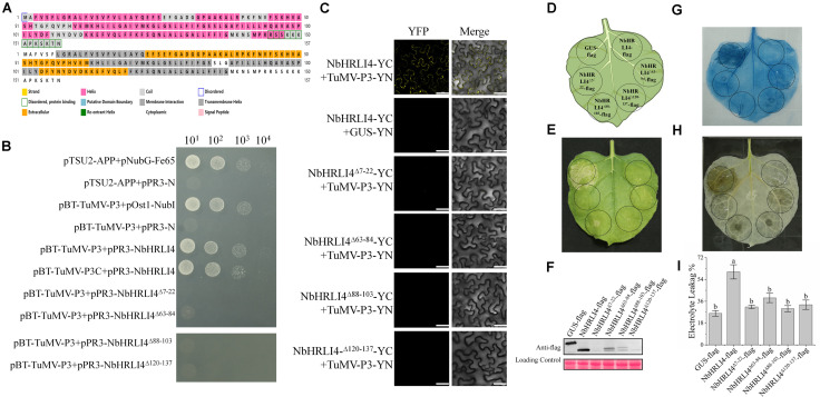
FIGURE 8.
The membrane interaction domains of NbHRLI4 are essential for its interaction with TuMV-P3 and for inducing cell death. (A) Protein structure analysis of NbHRLI4. (B) Y2H assays between pBT-TuMV-P3/pPR3-NbHRLI4, pBT-TuMV-P3C/pPR3-NbHRLI4, pBT-TuMV-P3/pPR3-NbHRLI4Δ7–22, pBT-TuMV-P3/pPR3-NbHRLI4Δ63–84, pBT-TuMV-P3/pPR3-NbHRLI4Δ88–103, and pBT-TuMV-P3/pPR3-NbHRLI4Δ120–137. pBT-TuMV-P3 with pPR3-N was used as negative control and pBT-TuMV-P3 with pOst1-NubI was the positive control. (C) BiFC assays were conducted between NbHRLI4/TuMV-P3, NbHRLI4Δ7–22/TuMV-P3, NbHRLI4Δ63–84/TuMV-P3, NbHRLI4Δ88–103/TuMV-P3, and NbHRLI4Δ120–137/TuMV-P3. GUS was used as negative control. Bars = 50 μm. (D) Schematic diagram of transient infiltration of GUS-flag, NbHRLI4-flag, NbHRLI4Δ7–22-flag, NbHRLI4Δ63–84-flag, NbHRLI4Δ88–103-flag, and NbHRLI4Δ120–137-flag in panels (E–G). (E) Overexpression of GUS-flag, NbHRLI4-flag, NbHRLI4Δ7–22-flag, NbHRLI4Δ63–84-flag, NbHRLI4Δ88–103-flag, and NbHRLI4Δ120–137-flag in bright at 6 dpi. (F) Western blotting detection of proteins expressed on leaves shown in panel (D). (G) Trypan blue staining of leaves expressing the vectors in panel (E) at 6 dpi. (H) DAB staining of leaves expressing the vectors in panel (E) at 6 dpi. (I) Leaf disks expressing the vectors in panel (E) were excised and assayed for electrolyte leakage at 6 dpi. The mean expression values were analyzed using F-test. Different letters on histograms indicate significant differences (P < 0.05).

