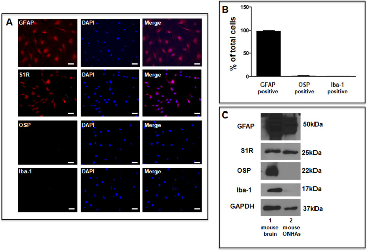
Figure 1.
Characterization of cultured primary mouse ONHAs. (A) ONHAs were fixed and probed with antibodies against GFAP (red), S1R (red), OSP (red), and Iba-1 (red). The cells were counterstained with DAPI to label DNA (blue) as a marker for nuclei. Scale bar: 50 µm. (B) Quantitative analysis shows that more than 95% of the cells in culture express GFAP. (C) The cell lysates from ONHAs (lane 2) were positive for GFAP, a marker for astrocytes and S1R, but negative for Iba-1, a marker for microglial cells, and OSP, a marker for oligodendrocytes. The protein extract from mouse brain (lane 1) was used as positive controls. Analyses were repeated in duplicate with cells isolated from different dates, different animals and treated on different days. For each immunofluorescence evaluation, three coverslips were quantified per group. Iba-1 is a marker for microglial cells.

