Figure 2.
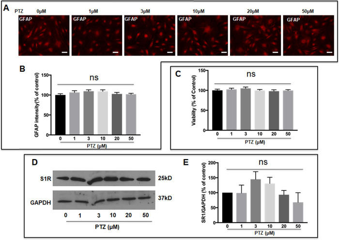
Effect of (+)-PTZ on WT ONHA viability, GFAP and S1R expression. WT ONHAs were treated with (+)-PTZ at varying concentrations (1, 3, 10, 20, or 50 µM) for 24 hours. (A) Representative images of GFAP expression detected by immunocytochemistry using GFAP antibody. Scale bar: 50 µm. (B) Quantification of GFAP staining intensity showed no significant differences with varying (+)-PTZ concentrations. (C) MTT assay showed no significant differences in ONHA viability with varying (+)-PTZ concentrations. (D) S1R expression was detected by Western blot and (E) quantified relative to GAPDH. No significant differences in S1R expression with varying (+)-PTZ concentrations were detected. Analyses were repeated in triplicate with cells isolated from different dates, different animals and treated on different days. For GFAP expression experiments, three coverslips were quantified from each group of each isolation. Eight microscopic fields were quantified per coverslip.

