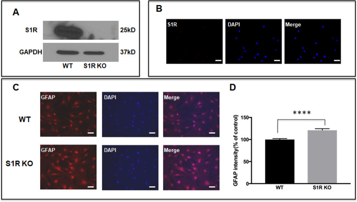
Figure 3.
GFAP expression in Sigma 1 Receptor knock out (S1R KO) ONHAs. (A) Western analysis showed that ONHAs isolated from S1R KO mice lacked S1R. (B) Immunocytochemistry showed that ONHAs isolated from S1R KO mice lacked S1R. Both WT and S1R KO ONHAs were plated on coverslips and incubated for 24 hours. (C) Representative images show cells stained with GFAP antibody. Scale bar: 50 µm. (D) S1R KO ONHAs expressed higher GFAP than WT ONHAs under the same cell culture conditions. These results were repeated five times with cells isolated from different dates, different animals and treated on different days. For each group of each isolation, three coverslips were quantified. Significantly different from control ****P < 0.0001. Data were analyzed using Student's t-test.

