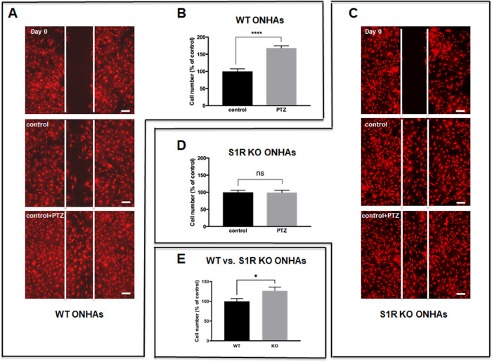
Figure 4.
Effect of (+)-PTZ on WT and S1R KO ONHA migration. WT and S1R KO ONHAs were pretreated with 10 µM (+)-PTZ or vehicle for 1h before scratching. After scratching, the cells were incubated with 10 µM (+)-PTZ or vehicle for 24 hours. (A) Representative images show the original scratch (day 0), and migration of WT ONHAs into the wound area after 24 hours of incubation with (+)-PTZ treatment or vehicle control. Scale bar: 200 µm. (B) Quantification of the number of WT ONHAs migrated into the wounded (scratched) area. Cells in wounded area were counted using ImageJ. For each group, four coverslips were quantified. WT ONHA migration increased significantly when cells were treated with (+)-PTZ. Significantly different from control ****P < 0.0001. (C) Representative images show the original scratch (day 0) and migration of KO ONHAs into the wound area after 24 hours of incubation with (+)-PTZ treatment or vehicle control. Scale bar: 200 µm. (D) Quantification of the number of KO ONHAs migrated into the wounded (scratched) area. Cells in wounded area were counted by ImageJ. For each group, three coverslips were quantified. (+)-PTZ did not affect S1R KO ONHA migration. (E) S1R KO control ONHAs had significantly more migrated cells than WT control ONHAs. Significantly different from control *P < 0.05. Data were analyzed using Student's t-test. These results were repeated in triplicate with cells isolated from different dates, different animals and treated on different days. For each group of each isolation, three coverslips were quantified.

