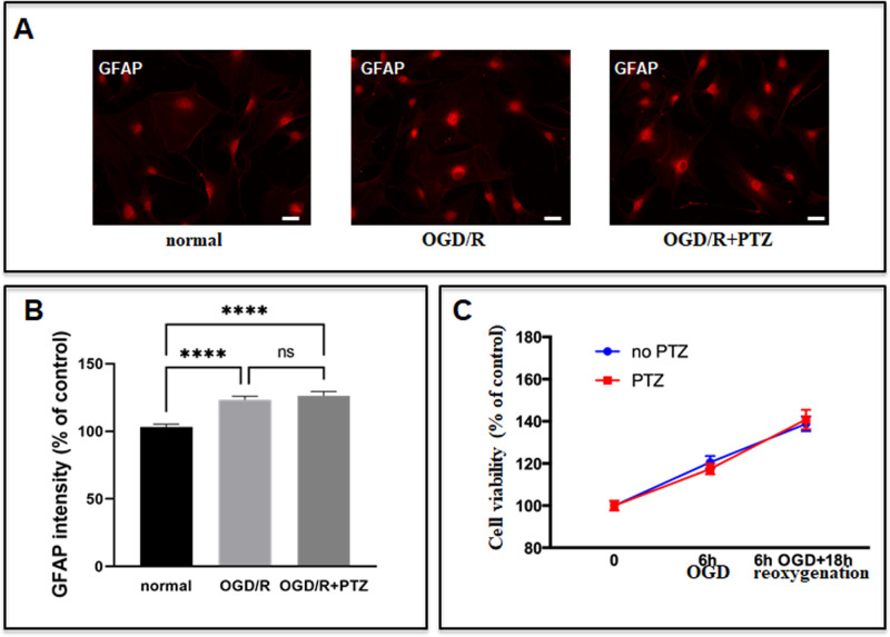
Figure 7.
Effect of (+)-PTZ treatment on OGD/R-exposed S1R KO ONHAs. (A) Representative images show GFAP expression in S1R KO ONHAs under normal and OGD/R-exposed conditions with or without (+)-PTZ treatment. Scale bar: 50 µm. (B) Quantitative analysis shows that OGD/R increases GFAP expression, and that (+)-PTZ treatment does not significantly enhance the OGD/R-induced increase in GFAP expression. Significantly different from control ****P < 0.0001, ***P < 0.001, *P < 0.05. (C) Cell proliferation was detected by MTT assay. Under conditions of OGD/R exposure, (+)-PTZ treatment did not significantly affect the S1R KO ONHA proliferation rate. Data were analyzed using two-way ANOVA followed by Tukey-Kramer post hoc test for multiple comparisons. These experiments were repeated in triplicate with cells isolated from different dates, different animals and treated on different days. For each group of each isolation, three coverslips were quantified.

