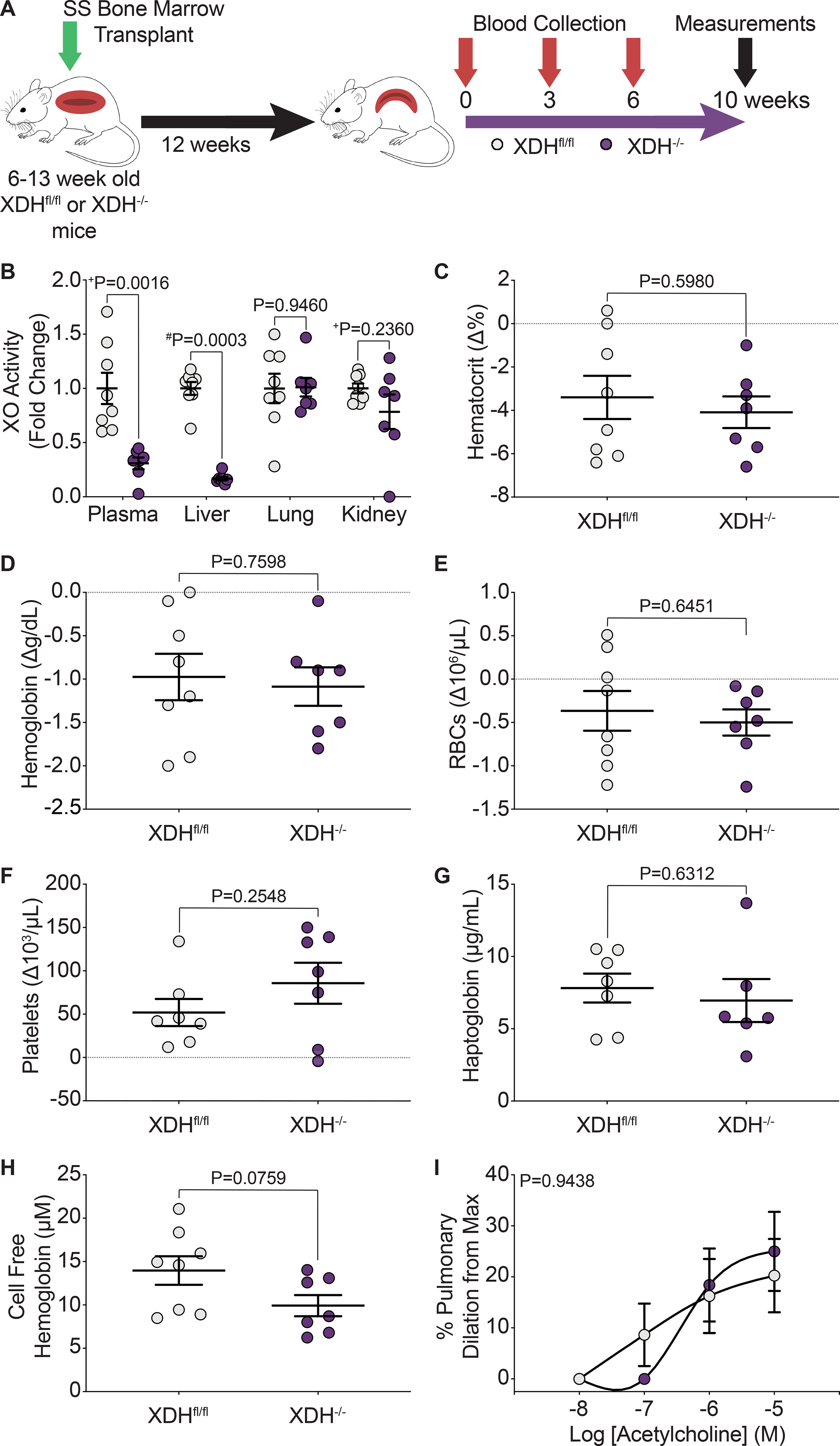
Figure 6. Hepatocyte-specific XO knockout did not decrease hemolysis or alter pulmonary vasoreactivity.

A) Experimental design. B) HPLC was used to measure plasma, liver, lung, and kidney XO activity. Complete blood counts show the hematological parameters C) hematocrit, D) hemoglobin, E) RBCs, and F) platelets, as a delta change from 0 to 10 weeks post-engraftment. G) An ELISA was used to measure plasma haptoglobin concentration 10 weeks post-engraftment. H) UV-visible spectral deconvolution was used to measure plasma cell free hemoglobin, a combination of oxyhemoglobin and methemoglobin, 10 weeks post-engraftment. Values are mean ± SEM using an unpaired Student’s t test unless otherwise noted. +Values are mean ± SEM using an unpaired Student’s t test with Welch’s correction. #Values are mean ± SEM using a Mann-Whitney test. I) An acetylcholine dose response was used to measure dilation of pulmonary arteries 10 weeks post-engraftment (Xdhfl/fl n=8, Xdh−/− n=7). Values are mean ± SEM using a two-way ANOVA with Sidak’s multiple comparisons test. XDH, xanthine dehydrogenase; XO, xanthine oxidase; RBCs, red blood cells; max, maximum; HPLC, high performance liquid chromatography ELISA, enzyme linked immunosorbent assay.
