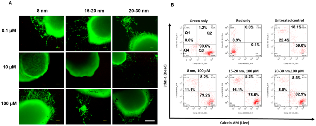
Figure 3.

Cell viability of cortical spheroids after the addition of iron oxide nanoparticles. (A) Live/Dead staining images for replated cortical spheroids after addition of iron oxide nanoparticles for 48 h. Scale bar: 200 μm. (B) Two-color flow cytometry dot plots for Live/Dead staining for the replated cortical spheroids after addition of iron oxide nanoparticles. The green only and red only controls indicate the proper color compensation.
