
Figure 4.
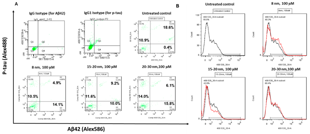
p-tau, Aβ42, and oxidative stress characterizations of cortical spheroids after addition of iron oxide nanoparticles. (A) Two-color flow cytometry dot plots for p-tau and Aβ42 expression for the replated cortical spheroids after addition of iron oxide nanoparticles for 48 h. (B) Flow cytometry analysis of reactive oxygen species for the replated cortical spheroids after addition of iron oxide nanoparticles for 48 h.