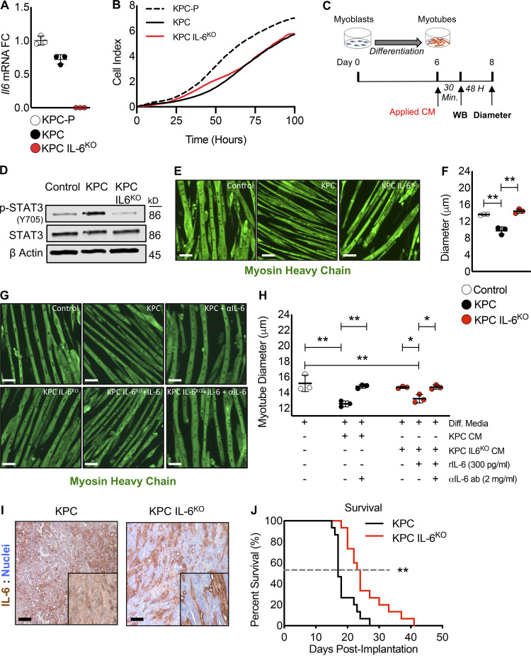
Figure 2.
Deletion of IL-6 from KPC cells prevented muscle wasting in vitro and increased survival in mice. (A) Targeted mutagenesis of the Il6 gene was performed, and a transfection control clone (KPC) and an Il6 ablated clone (KPC IL6KO) were selected for use in downstream experiments based on their Il6 expression relative to the untransfected parental cell line (KPC-P). Clones were cultured individually in triplicate for n = 3 per group, and error bars are mean plus SD. (B) To determine if deletion of IL-6 affected tumor cell growth, a proliferation assay was performed comparing the clones, where clones were cultured in triplicate (n = 3 per group) and measurements were taken every hour for 100 h; growth curves represent the mean proliferation of triplicate wells over time. (C) Myoblasts were differentiated into myotubes and treated in triplicate (n = 3 per group) with 30% CM from tumor clones to measure effects on myotube atrophy and the activation of STAT3. (D) Western blotting (WB) analysis using the pooled myotube protein lysates from n = 3 per group was performed to measure phosphorylation of STAT3 (p-STAT3) as an indication of STAT3 activation. (E and F) Myotubes were visualized using IF with an anti-myosin heavy chain (MHC) antibody (E), and myotube atrophy was measured from 20 random fields per well (n∼150 myotube diameter measurements per well, n = 3 wells per group). Scale bar = 50 µm. Error bars represent mean myotube diameter and SD, and significant differences (**, P < 0.001) were determined using ANOVA and Tukey’s multiple comparisons test (F). (G and H) To verify atrophy was influenced by IL-6, myotubes were treated in triplicate with KPC CM and IL-6 neutralizing antibody as well as KPC IL6KO CM plus recombinant IL-6 with and without the presence of an anti–IL-6 neutralizing antibody. Myotubes were visualized with MHC IF, and diameter was measured as described in F. Scale bar = 50 µm. Error bars are mean and SD, and significant differences (*, P < 0.05; **, P < 0.001) were determined using ANOVA and Tukey’s multiple comparisons test. (I) KPC tumor cells were orthotopically implanted into mice, and tumors were excised, sectioned, and reacted with anti–IL-6 IHC to verify tumor cell IL-6 deletion. Insets are increased magnification to show staining; scale bar = 20 µm. (J) Survival was measured in mice orthotopically implanted with KPC and KPC IL6KO tumor cells. The dashed line represents median survival, n = 15 per group, and statistical difference (**, P < 0.001) was determined using a Kaplan-Meier estimate. All panels represent data from single experiments. Diff. media, DM.

