Figure 3.
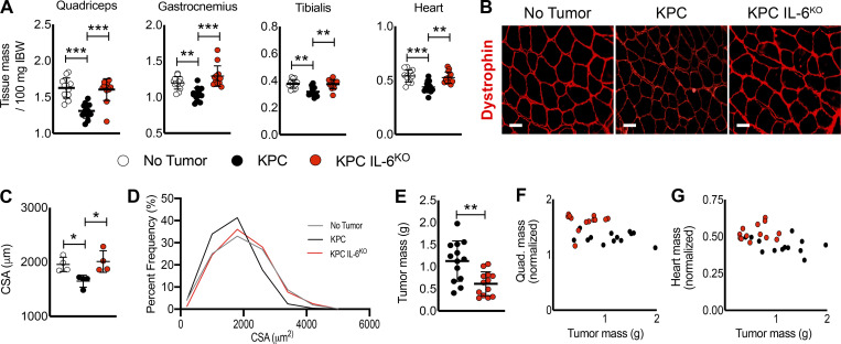
Deletion of tumor cell IL-6 attenuates muscle wasting. (A) KPC tumor–bearing mice reached our humane endpoint criteria 17 d after injection, and all groups were euthanized simultaneously. Skeletal muscles and the heart were excised at euthanasia and weighed and normalized to initial body weight (IBW). Data represent the mean plus SD from two individual experiments (n = 13 per group); significant differences (**, P < 0.005; ***, P < 0.0005) were determined using ANOVA and Tukey’s multiple comparisons test. (B and C) Evaluation of muscle fiber CSA was done using sections from excised quadriceps muscles from individual mice reacted for dystrophin expression (B), and mean fiber CSA was determined by comparing myofiber means from muscle cross sections from a subset of samples (n = 4; n > 300 myofibers per animal), quantified from 20 random fields from each cross section (C). Scale bar = 50 µm. Error bars are mean and SD, and significant differences (*, P < 0.05) were determined using ANOVA and Tukey’s multiple comparisons test. (D) Cumulative fiber CSA values from mice in each group were organized based on percent distribution of fiber CSA and plotted to observe shifts in distribution. (E) Tumors were excised, and tumor mass was recorded. Data represent the mean plus SD from two individual experiments (n = 13 per group); significant differences (**, P < 0.005) were determined using Student’s t test. (F and G) Correlation analysis of tumor size and muscle size for the quadriceps and heart showed no correlations.

