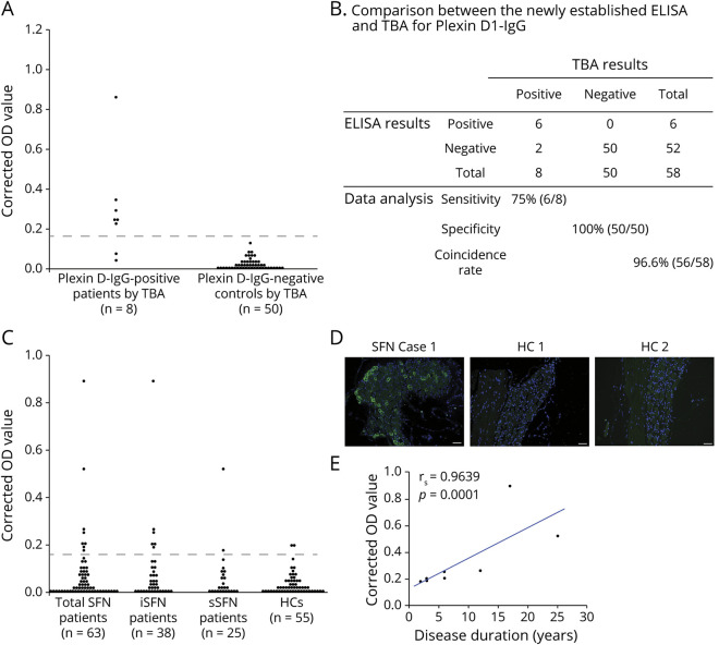
Figure 1. ELISA Screen and Confirmatory TBA for Plexin D1-IgG.
(A) Indirect ELISA for plexin D1-IgG in a previous cohort of 8 patients with NeP with plexin D1-IgG and 50 non-NeP patients (30 disease controls and 20 HCs) previously determined by TBA. Disease controls included 6 with amyotrophic lateral sclerosis; 4 each with multiple system atrophy, systemic lupus erythematosus, and neuro-Behçet disease; 3 with hereditary spinocerebellar degeneration; 2 each with Parkinson disease, normal pressure hydrocephalus, and Sjögren syndrome; and 1 each with Alzheimer disease, dementia with Lewy bodies, and corticobasal degeneration. The difference in OD values between plexin D1-coated wells and D1-uncoated wells (corrected OD value) was calculated, and the test was considered “ELISA-positive” when the corrected OD value was above the mean + 5 SD of the 50 plexin D1-IgG-negative controls determined by TBA (0.163, dotted line). Six of 8 patients with NeP with plexin D1-IgG by TBA were positive for plexin D1-IgG by ELISA, whereas all 50 disease controls and HCs without plexin D1-IgG by TBA were negative for plexin D1-IgG by ELISA. (B) Comparison of the newly established ELISA with TBA for plexin D1-IgG. The overall coincidence rate of ELISA to TBA was 96.6% (56/58). (C) Indirect ELISA for plexin D1-IgG in the present SFN cohort. The difference in OD values between plexin D1-coated wells and D1-uncoated wells (corrected OD value) was calculated, and the test was considered “ELISA-positive” when the corrected OD value was above 0.163 (dotted line). Plexin D1-IgG was positive in 8 of 63 (12.7%) of all patients with SFN, including 6 of 38 (15.8%) patients with iSFN, 2 of 25 (8.0%) patients with sSFN, and 2 of 55 (3.6%) HCs by ELISA. (D) IgG (green) from a representative patient with SFN (iSFN Case 1 in table 2) showed positive immunostaining of mouse small DRG neurons, whereas there was no significant immunoreactivity in 2 ELISA-seropositive HCs (HC 1 and HC 2). Nuclei are counterstained with 4′,6-diamidino-2-phenylindole (DAPI) (blue). (E) Correlation between the corrected OD value and the disease duration in patients with SFN with plexin D1-IgG (n = 8). There was a significant positive correlation between them (Spearman rank correlation; rs = 0.9639, p = 0.0001). Even after 1 outlier with the highest optical density was removed, the correlation between the corrected OD value and the disease duration remained significant (rs = 0.982, p < 0.0001). HC = healthy control; IgG = immunoglobulin G; iSFN = idiopathic small fiber neuropathy; OD = optical density; SFN = small fiber neuropathy; sSFN = secondary small fiber neuropathy; TBA = tissue-based indirect immunofluorescence assay.

