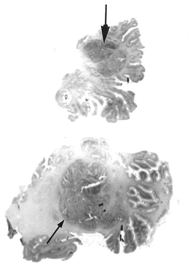
Fig 2.
Coronal microscopic sections (posterior to anterior view) of the cerebellar lesions at vermian (top) and hemispheric (bottom) levels show vermian hypoplasia with irregularly shaped foliation. A large central multinodular conglomerate (arrows) corresponding to juxtaposed ectopic polymicrogeria nodules is seen in the right hemisphere and vermis.

