
Fig 2.
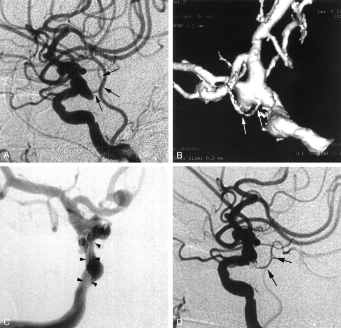
Ruptured internal carotid artery aneurysm.
A, Lateral 2D-DSA image. A wide-necked aneurysm can be seen at the origin of the posterior communicating artery (arrows). The relationship of the aneurysm to the posterior communicating artery is not clear in this image.
B, Surface shaded display image. Note the neck of the aneurysm and its involvement with the posterior communicating artery (arrows). The dome-to-neck ratio is 1.0 on this three-dimensional image. The calculated difficulty score is 3.
C, GDC procedure was performed with a double microcatheter (arrowheads) technique.
D, Lateral 2D-DSA image obtained immediately after GDC procedure shows incomplete occlusion (<95%) of the aneurysm and patent posterior communicating artery (arrows).