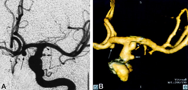
Fig 1.
Patient 12, aneurysm 21. Example of smallest aneurysm seen in this study.
A, Midarterial phase anteroposterior projection DSA image shows a small saccular aneurysm at the supraclinoid internal carotid artery (short arrow). Note larger anterior communicating artery aneurysm (long arrow). Anterior choroidal artery aneurysm is mostly obscured by overlying internal carotid artery bifurcation (arrowhead).
B, 3D posteroanterior projection CTA image with lateral angulation shows aneurysm sac (short arrow). Anterior communicating artery (long arrow) and anterior choroidal artery (arrowhead) aneurysms are also well seen.

