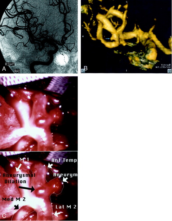
Fig 2.

Patient 14, aneurysm 25. Example of DSA false negative finding due to overlapping MCA branches.
A, Right internal carotid artery injection, midarterial phase right anterior oblique projection (one of many different projections obtained) DSA image fails to show vascular abnormality at right MCA bifurcation.
B, Volume-rendered 3D direct inferosuperior projection CTA image shows a 2.7-mm aneurysm of the right MCA bifurcation with laterally projecting sac (arrow).
C, Intraoperative photograph without (top) and with (bottom) labels, with sylvian fissure retracted, shows small laterally projecting saccular aneurysm of the left MCA bifurcation (arrow) and small placoid aneurysmal dilation, not well seen on CTA images. Ant, anterior; Temp, temporal; Med, medial; Lat, lateral.
