Fig 3.
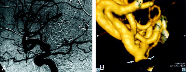
Patient 18, aneurysm 32. Example of CTA false negative (reader 1) finding.
A, Midarterial phase right anterior oblique projection DSA image shows a small pyramidal aneurysm sac projecting inferiorly from the anterior M2 division (straight arrow). Note the small anterior temporal branch arising from the neck region (curved arrow).
B, 3D lateromedial projection CTA image clearly shows the pyramidal aneurysm sac (straight arrow), along with a 0.5-mm anterior temporal branch arising from the region of the aneurysm neck (curved arrow). The aneurysm is clearly present but was overlooked during the initial reading by reader 1.

