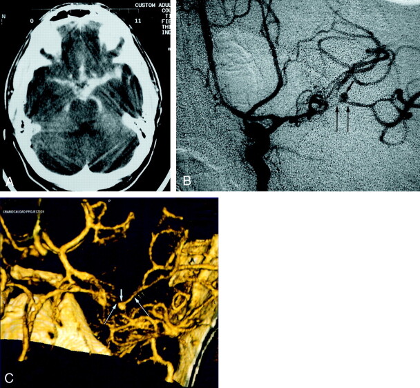
Fig 4.
Patient 6, aneurysm 9. Example of ability of CTA to visualize small aneurysms in the setting of acute severe SAH.
A, Axial unenhanced CT scan shows extensive hyperattenuated blood in the sylvian and choroidal fissures and in the basal cisterns.
B, Midarterial phase anteroposterior projection DSA image of the left internal carotid artery shows a small saccular aneurysm at the MCA bifurcation (short arrow) and severe vasospasm of the distal M1 and proximal M2 segments (long arrows).
C, Volume-rendered 3D CTA image shows a 4.0-mm maximal diameter saccular aneurysm of the left MCA bifurcation (short arrow) and also luminal reduction compatible with severe segmental vasospasm of the distal left M1 and proximal M2 segments (long arrows).

