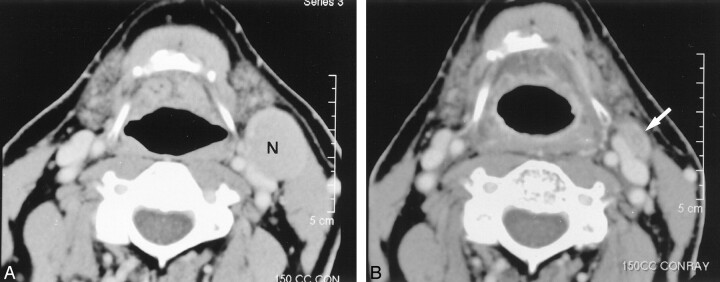
Fig 2.
Images in a 64-year-old man with metastatic neck disease from an unknown primary squamous cell carcinoma (TXN2A).
A, Pre-RT contrast-enhanced CT image reveals an enlarged (27 × 22 mm) level 2 lymph node (N) on the left.
B, Post-RT contrast-enhanced CT image shows a small residual mass (arrow) (decrease ratio of the largest dimension of the node, 52%). The patient underwent left neck dissection after this study. The specimen from the left hemineck was negative.

