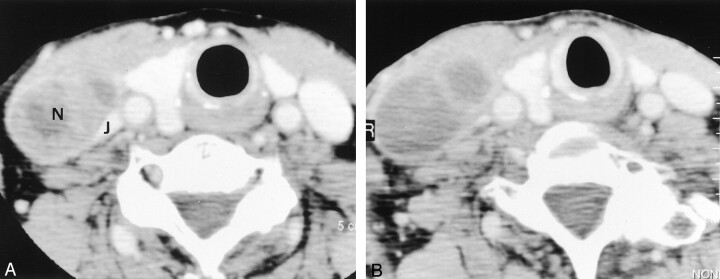
Fig 4.
Images in a 78-year-old woman with pyriform sinus carcinoma (T2N2A).
A, Pre-RT contrast enhanced CT image shows an enlarged (40 × 25 mm) level 4 lymph node (N) on the right. The nodal mass distorts the adjacent right internal jugular vein (J).
B, Post-RT contrast-enhanced CT image shows a slight increase in the size of the lymph node (decrease ratio of the largest dimension, −20%). The internal attenuation is generally lower on this image than on the image in A. The specimen from the right hemineck was positive.

