Fig 5.
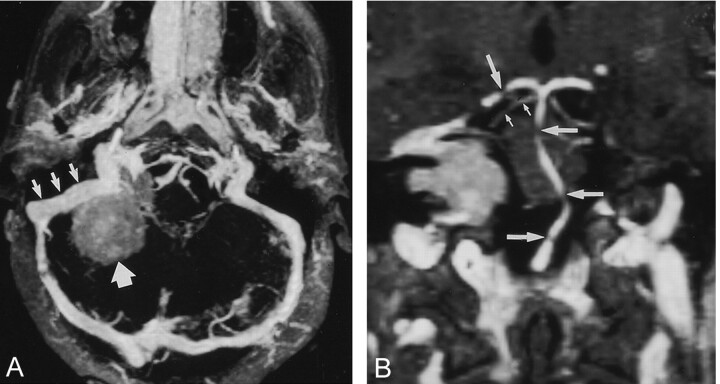
Images in a 42-year-old patient with meningioma in right cerebellopontine angle.
A, Real-time para-axial MIP image with 15-mm thickness shows the sigmoid sinus (small arrows) in relation to a meningioma (large arrow), which has signal intensity lower than that of the sinus. No signs of tumor infiltration are depicted.
B, Real-time paracoronal MIP image with 2-mm thickness shows the relation of the posterior fossa arteries to the tumor. Gaps in the basilar artery (large horizontal arrows) and in the right posterior cerebral artery (large vertical arrow) are due to partial volume effects. The proximal superior cerebellar artery is shown with great detail (small arrows).

