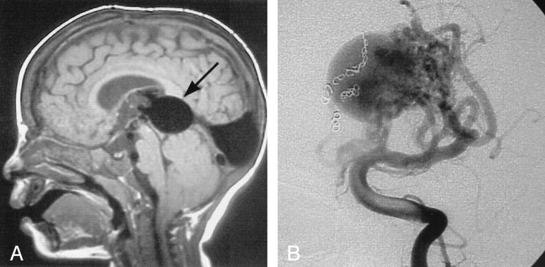
Fig 1.
Images from the case of a 19-month-old patient with mild hydrocephalus and engorged scalp veins.
A, Sagittal view T1-weighted MR image shows the markedly enlarged median prosencephalic vein of Markowski, characteristic of VGAM (arrow). Arterial feeders can be seen along the anterior wall of the vein.
B, The complex arterial maze (arrows) is well seen on this conventional angiogram obtained with injection of the left vertebral artery. Coils can be seen along the right side of the varix, occluding several arterial feeders.

