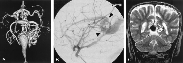
Fig 2.
Images from the case of a 3-month-old triplet with worsening congestive heart failure.
A, Volume-rendered MR angiogram shows, from an inferior prospective, the arterial feeders supplying the lesion from the left side (arrows).
B, Carotid injection during arterial embolization procedure performed when the patient was 6 months old shows residual flow to the lesion from pericallosal and posterior choroidal arteries (arrowheads).
C, Coronal view fast spin-echo T2-weighted MR image obtained 6 years after embolization shows continued patency of the varix and mild enlargement of extra-axial fluid spaces.

