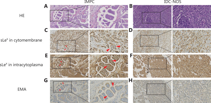
Figure 1.
Immunohistochemical expression of sLex and MUC1/EMA in breast IMPC and IDC-NOS. (A) IMPC consists of tumor cell clusters of epithelial cells surrounded by gaps [hematoxylin and eosin (HE) staining]. (B) Representative microphotographs of IDC-NOS (HE). Immunohistochemistry confirming the expression of sLex on the cytomembranes in IMPC cell clusters (C) and IDC-NOS cells (D). The expression of sLex within the cytoplasm in IMPC cell clusters (E) and IDC-NOS cells (F). EMA immunostaining showing the typical polarity reversal growth pattern of IMPC (G) and the normal pattern in IDC-NOS tumor cells (H) (200×, 400×, indicated by the red arrow).

