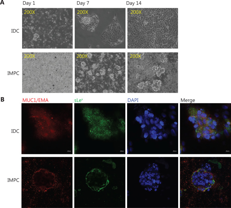
Figure 2.
Immunofluorescence expression of sLex and MUC1/EMA in breast IMPC and IDC-NOS. (A) Cell morphology of primary cultured IMPC and IDC-NOS cells. Primary IMPC cells are suspended, non-adherent tumor cell clusters; primary IDC cells are adherent tumor cells (200×). (B) Immunofluorescence confirming the expression of MUC1/EMA and sLex on the cytomembranes in IMPC cell clusters and IDC-NOS cells (630×).

