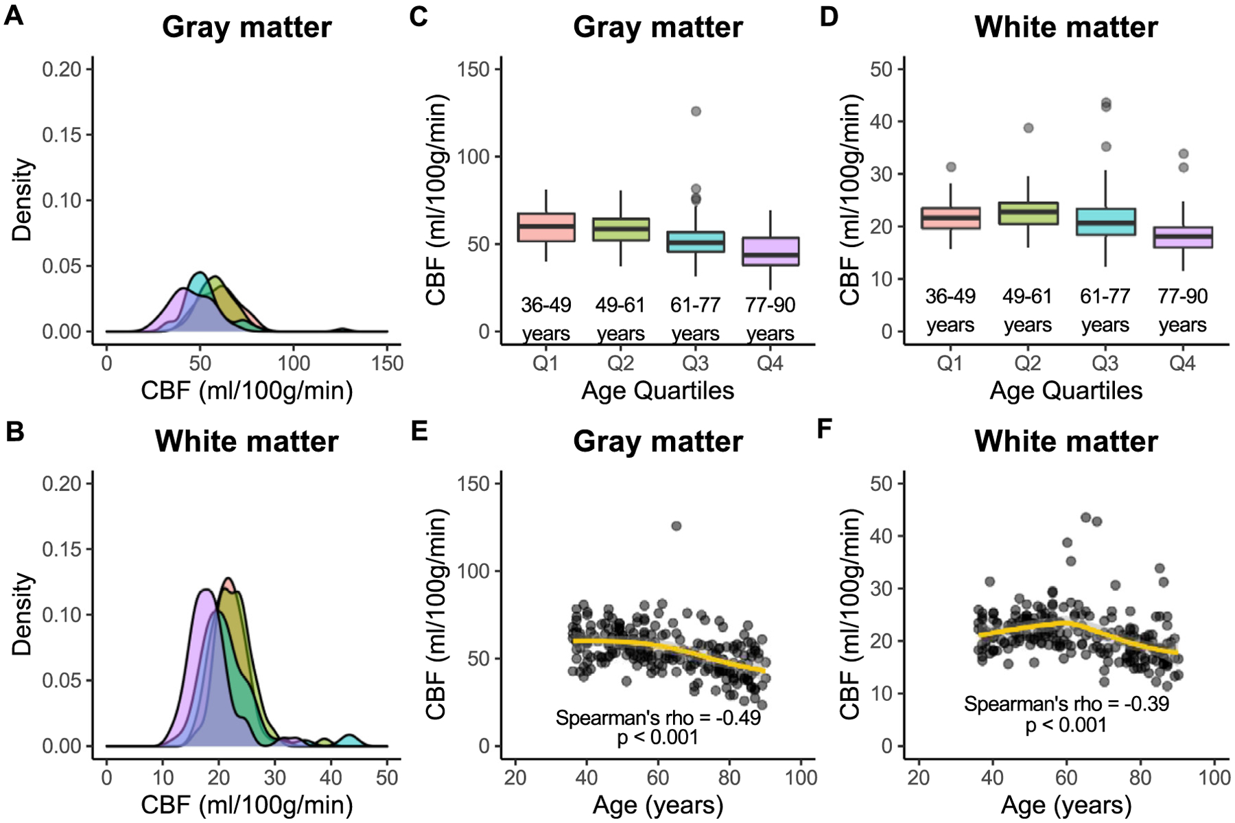
Fig. 3. Cerebral blood flow.

Associations between cerebral blood flow (CBF) and age are shown here in gray and white matter across quartiles of age: Q1 (36–49 years; red), Q2 (49–61 years; green), Q3 (61–77 years; blue), and Q4 (77–90 years; purple). Kernel density plots show that the distribution of gray matter CBF (A) is wider than that of white matter (B) in all age groups. In addition, gray matter CBF (C) is significantly higher than white matter CBF (D) in all age groups. Finally, CBF is inversely associated with age in both gray matter (E) and white matter (F), but this relationship is stronger in gray versus white matter.
