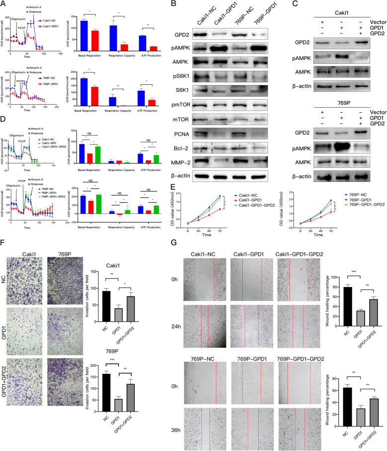
Fig. 3.
Overexpression of GPD1 inhibited mitochondrial function and induced AMPK phosphorylation by suppressing the expression of GPD2. (A) Oxygen consumption rate (OCR) of Caki1 and 769P cells with GPD1 overexpression. Baseline respiration, spare respiratory capacity, and ATP production in Caki1 and 769P cells with GPD1 overexpression. (B) Western blotting of indicated proteins in Caki1-NC, Caki1-GPD1, 769P -NC, and 769P-GPD1 cells. (C) Western blotting of GPD2, pAMPK, and AMPK in Caki1-GPD1 and 769P-GPD1 cells with GPD2 restoration and a negative control. (D) The OCR in Caki1-GPD1 and 769P-GPD1 cells with GPD2 restoration and a negative control. Baseline respiration, spare respiratory capacity, and ATP production in Caki1-GPD1 and 769P-GPD1 cells with GPD2 restoration and a negative control are shown. (E) Cell proliferation in Caki1-GPD1 and 769P-GPD1 cells with GPD2 restoration and a negative control as examined by CCK-8 assays. (F–G) Cell migration and invasion in Caki1-GPD1 and 769P-GPD1 cells with GPD2 restoration and a negative control as examined by wound healing and Transwell assays. Red lines denote the margins of the wound. Data are shown as the mean + SD (n = 3). Statistical analysis was performed using an unpaired Student’s t test with a two-tailed distribution (NS, p > 0.05, * p < 0.05, **p < 0.01, ***p < 0.001). Abbreviations: GPD1, glycerol-3-phosphate dehydrogenase 1; GPD2, glycerol-3-phosphate dehydrogenase 2; CCK-8, cell counting kit

