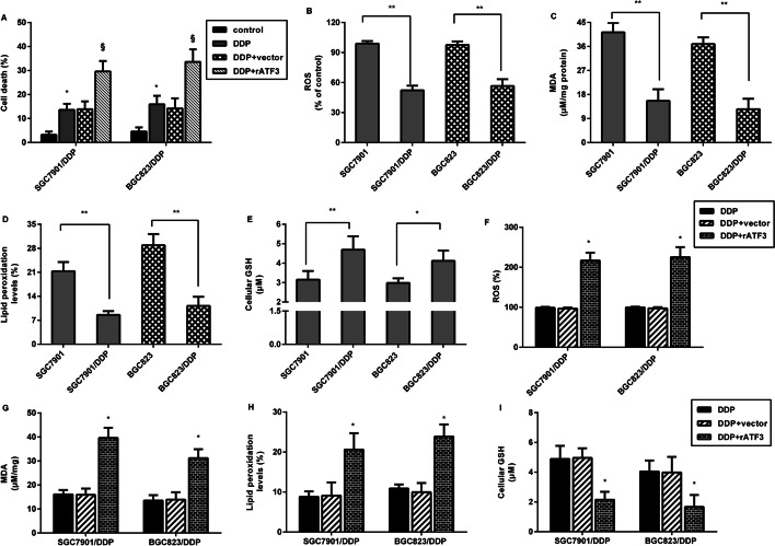
Fig. 3.
ATF3 evoked ferroptosis in GC cells. A SGC7901/DDP and BGC823/DDP cells transfected with ATF3 plasmids were exposed to cisplatin. Cell death was then assessed by Annexin V-FITC/PI. B–E Ferroptosis was evaluated in cisplatin-resistant and parental GC cells by detecting ROS (B), MDA (C), lipid peroxidation (D) and GSH levels (E). F–I Cisplatin-resistant GC cells were treated with ATF3 vector transfection and cisplatin exposure, and the ROS (F), MDA (G), lipid peroxidation (H) and GSH levels (I) were then measured. *P < 0.05. **P < 0.01

