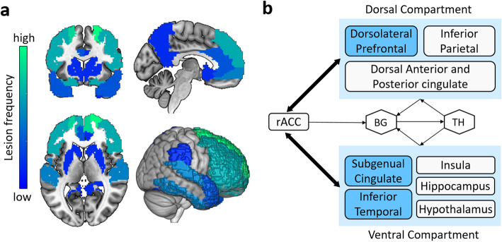
Fig. 3.
The brain lesion patterns and frontal-limbic circuit of depression in AD. a The brain region with the highest frequency of depression lesions is the superior frontal lobe, followed by the left inferior temporal and other frontal regions, as well as other temporal regions, the anterior cingulate, entorhinal, right hippocampal, caudate nucleus, lentiform nucleus, fusiform, right posterior cingulate, precuneus, supramarginal and parietal lobe. b The frontal-limbic circuit is composed of a dorsal part dominated by the dorsolateral prefrontal cortex and ventral part dominated by the subgenual cingulate and inferior temporal cortex. A direct projection from the subgeniual cingulate to the dorsolateral prefrontal cortex and a bidirectional indirect pathway through multiple marginal regions, including the posterior cingulate, hypothalamus, hippocampus, and insula are delineated. Abbreviations: rACC = rostral anterior cingulate; BG = basal ganglia; Th = thalamus. b A visual adaptation of a figure from Mayberg et al. [99], with permission

