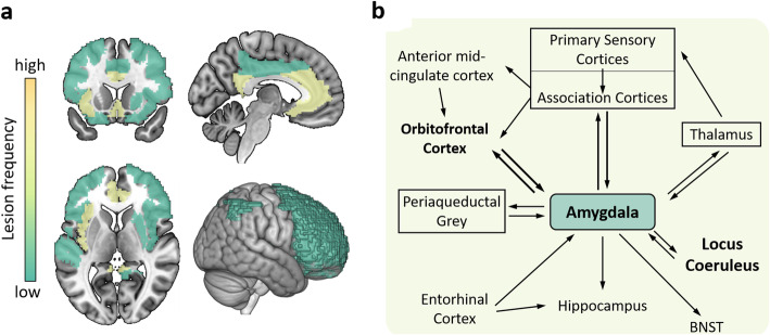
Fig. 4.
The brain lesion pattern and amygdala circuit of anxiety in AD. a The anterior and posterior cingulate cortex, entorhinal cortex, parahippocampal gyrus and insula cortex are the highest frequency of anxiety lesion regions, and the second is the amygdala, right precuneus, inferior parietal lobule, left anterior superior temporal, putamen, middle cingulate cortex and the frontal lobe. b The afferent arm of the anxiety circuit includes the exteroceptive sensory systems of the brain, which convey the sensory information contained in anxiety-inducing stimuli to the dorsal thalamus. An exception is the olfactory system, which carries information through the amygdala and entorhinal cortex, not the thalamus. Visceral afferent pathways alter the function of the locus coeruleus and amygdala. The thalamus relays sensory information to the primary sensory receptive areas of the cortex, which project to adjacent unimodal and polymodal cortical association areas. The cortical association areas send projections to the amygdala, entorhinal cortex, orbitofrontal cortex, and cingulate gyrus. The efferent pathways involving the amygdala, locus coeruleus, hypothalamus, periaqueductal gray, and striatum mediate autonomic, neuroendocrine, and skeletal-motor responses are associated with anxiety. Abbreviations: BNST = bed nucleus of the stria terminalis. b A visual adaptation of a figure from Charney et al. [100], with permission

