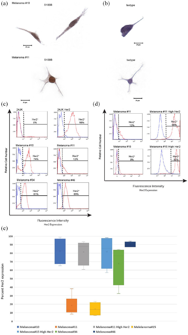
Figure 5.
Melanoma tumor cells constitutively express Her2 antigen.
(a-b) Melanoma tumor cells #10 and #11 were cultured onto slides and fixed using formaldehyde and stained for S100B. Images were taken using an Olympus BX51 Microscope at ×40 magnification and captured using SpotFire software. Single representative cells are presented stained with either an S100B-specific antibody (a) or isotype control (b). (c) Tumor lines were examined for Her2 antigen by flow cytometry. Cells were gated on morphology, viability and Her2 antigen expression. Cells that express Her2 antigen (red) are demonstrated against an isotype for Her2 (blue). 24JK tumor lines negative and positive for Her2 expression were used as additional controls. (d) Cell lines of melanoma tumors #11 and #15 were transduced using lipofectamine transduction protocols and sorted for Her2 expression. Control lines with 24JK parental and Her2 expressing tumor lines are shown. Melanoma tumor cell lines from patients #11 and #15 are shown before transduction (Melanoma#) and after transduction and sorting (Melanoma# High Her2). Her2 expressing cells are demonstrated in red and isotype staining is demonstrated in blue. (e) Box plot demonstrating range of Her2 antigen expression for each patient cell line (n = ⩾4 observations per patient).

