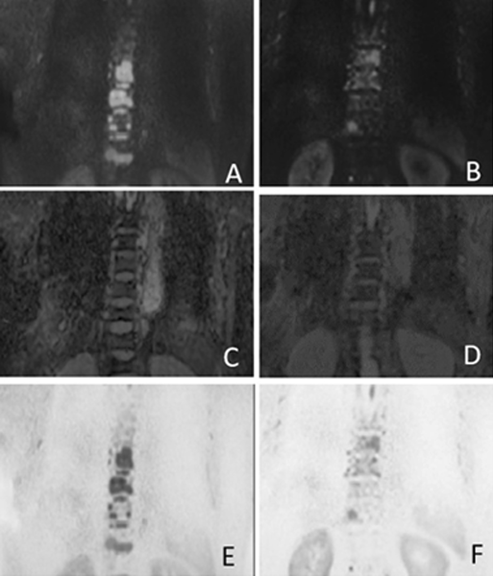
Fig. 1.

The patient underwent WB-DWI after the diagnosis of multiple myeloma. A (DWI b = 1000 s/mm2), C (ADC map) and E (inverted MIP image) show MM lesions of spine at baseline visit. B (DWI b = 1000 s/mm2), D (ADC map) and F (inverted MIP image) show MM lesions after 4-course chemotherapy. The ADC value of the lesion in the third lumbar vertebra increased from (0.698 × 10⁻³ mm2/s) to (0.796 × 10⁻³ mm2/s). In b = 1000 s/mm2 DWI images, the lesion area in B after treatment was significantly smaller than that in A before treatment. This contrast is more intuitive on the inverted image, which is shown in F after treatment and E before treatment
