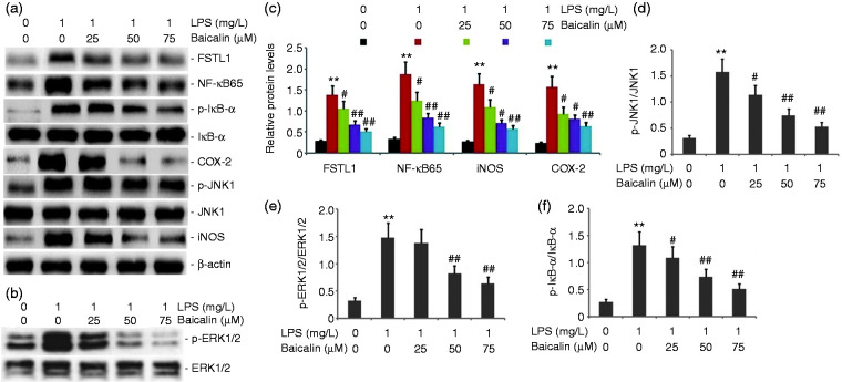
Figure 12.
Effect of baicalin on the activity of the FSTL1 and ERK/JNK signaling pathway in LPS-induced alveolar type II epithelial cells. A549 cells were incubated with LPS (1.0 mg/l) and various concentrations of baicalin (25, 50, and 75 μM) for 24 h. FSTL1, p-ERK1/2, p-JNK1, NF-κB65, p-IκB-α, iNOS, and COX-2 protein expression levels were measured by Western blot. The data from three independent experiments is presented as the mean ± SEM. **P < 0.01 versus the control group. #P < 0.05; ##P < 0.01 versus only LPS group. P < 0.05 versus the baicalin (25 μM) group versus the baicalin (50 μM) group and the baicalin (75 μM) group. P > 0.05, the baicalin (50 μM) group versus the baicalin (75 μM) group. a and b: Representative Western blots of FSTL1, p-ERK1/2, p-JNK1, NF-κB65, p-IκB-α, iNOS, and COX-2 protein expression in LPS-induced alveolar type II epithelial cells. c–f: Statistical summary of the densitometric analysis of FSTL1, p-ERK1/2, p-JNK1, NF-κB65, p-IκB-α, iNOS, and COX-2 protein expression in LPS-induced alveolar type II epithelial cells.

