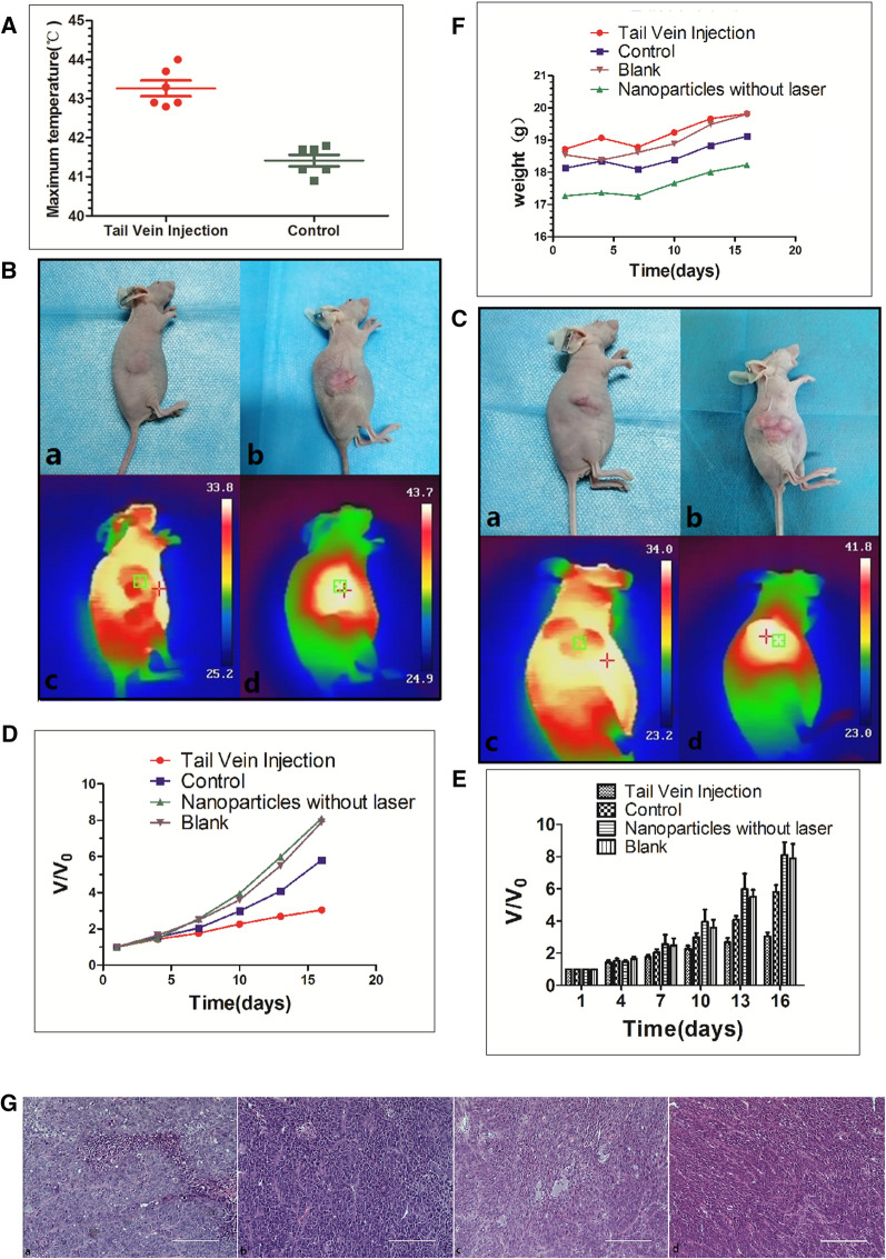
Fig. 7.
A The highest temperature of nude mice xenografts in different groups after 10 min of NIR light irradiation. B Tail vein injection group. "+" is the highest temperature area which measured by NIR imager. (a) Tumor before treatment, (b) Tumor after 5 treatments, (c) The maximum temperature is 33.8 °C (non-tumor tissue temperature) before treatment, (d) The maximum temperature after 10 min of treatment (tumor tissue temperature) reached 43.7 °C. C Control group. "+" is the highest temperature area which measured by NIR imager. (a) Tumor before treatment, (b) Tumor after treatment 5 times, (c) The highest temperature before treatment is 34.0 °C (non-tumor tissue temperature), (d) The highest temperature after treatment reaches 41.8 °C 10 min (tumor tissue temperature). D and E Changes in tumor volume pre- and post-treatment in the tail vein injection group, control group, nanoparticles without laser irradiation group and blank group. F Changes of weight in nude mice pre- and post-treatment among the tail vein injection group, control group, nanoparticles without laser irradiation group and blank group. G HE staining. More apoptotic cells were seen in the tail vein injection group (a), but very few in the control group (b), the blank group (c) and the nanoparticles without laser irradiation group (d)

