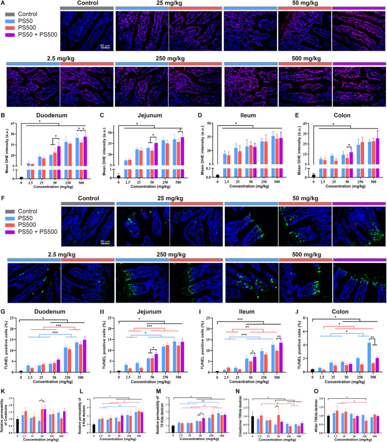
Fig. 7.
28-day repeated dose oral exposure to PS micro- and nanoplastics confirmed the combined effects. A Representative merged images of the jejunum sections stained with DHE to assess the ROS generation after repeated dose of PS particles exposure for 28 days. The ROS was stained in red and the nucleus was stained with DAPI in blue. The quantification of mean DHE intensity in B) duodenum, C) jejunum, D) ileum and E) colon was shown. F Representative merged images of the jejunum sections stained with TUNEL to assess the cell apoptosis after repeated dose of PS particles exposure for 28 days. The apoptotic cells were stained in green and the nucleus was stained with DAPI in blue. The quantification of TUNEL positive cell rate in G) duodenum, H) jejunum, I) ileum and J) colon was shown. The fluxes of K) creatinine, L) 4 kDa dextran, M) 70 kDa dextran in the intestine were quantified. The fluxes of N) creatinine and O) 4 kDa dextran were normalized to 70 kDa dextran flux. *P < 0.05. Results were shown as means ± SE. Comparisons were made with ANOVA, followed by Tukey’s method (n = 5 per group). The color bars were used for grouping. The exposure dose was shown by the color bars

