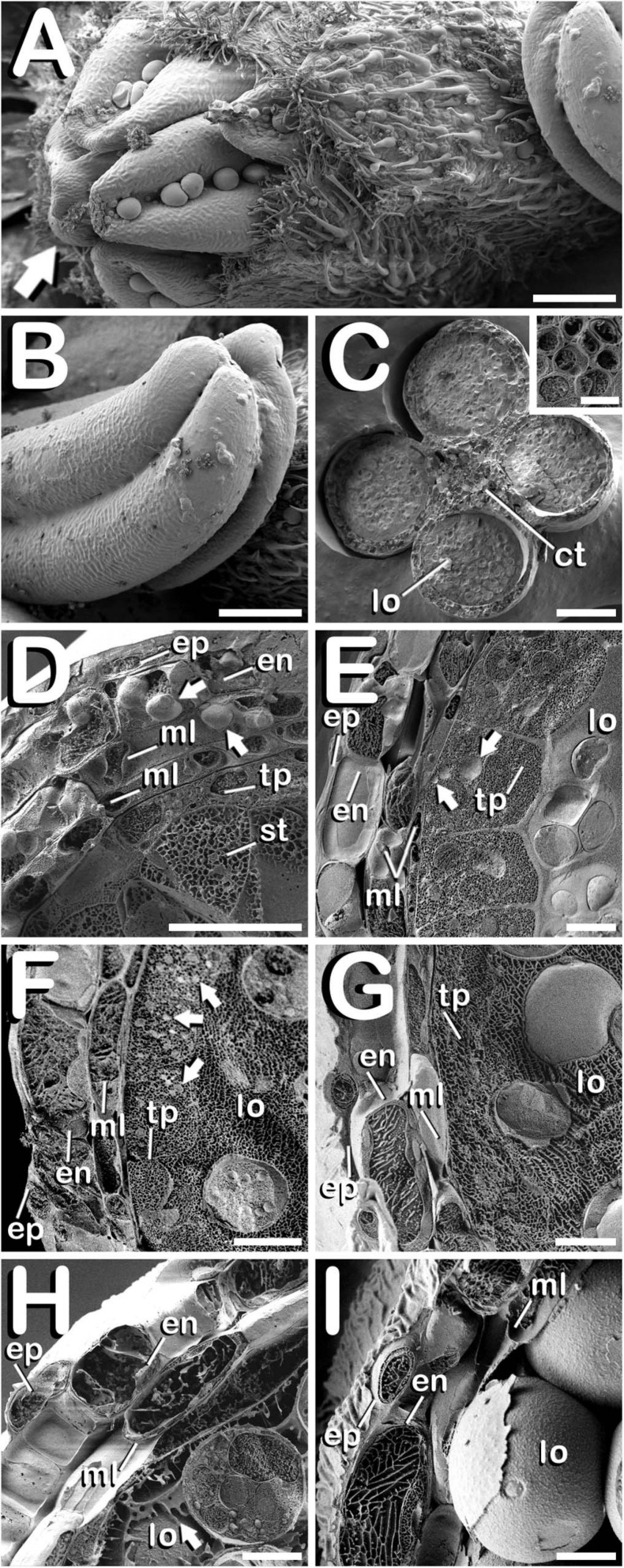
FIGURE 5.
Cryo-SEM images showing anther wall development in C. sativa. The different developmental stages are described as follows: (A) Staminate flower with its five anthers being visible: arrow points to the anthers. (B) Anther excised from a male bud. (C) Cross-section of an anther after freeze-fracture showing four microsporangia and vascular bundles from connective tissue (inset in Figure 5C). (D) Microspore mother cell stage: arrows point plastids in endothecium and outer middle layer. (E) Tetrad stage: arrows point orbicules embedded in tapetum layer. (F) Young and mid microspore stage: arrows point orbicules embedded in tapetum layer. (G) Vacuolate microspore stage. (H) Young bi-cellular pollen stage: arrow points to tapetum sticky remnants covering the pollen grain surface. (I) Mature pollen stage. Scale bars (A,B): 200 μm. Scale bars (C): 100 μm. Scale bar (inset in C): 5 μm. Scale bars (D–I): 10 μm. lo, locule; ct, connective tissue; ep, epidermis; en, endothecium; ml, middle layer; tp, tapetum; st, sporogenous tissue.

