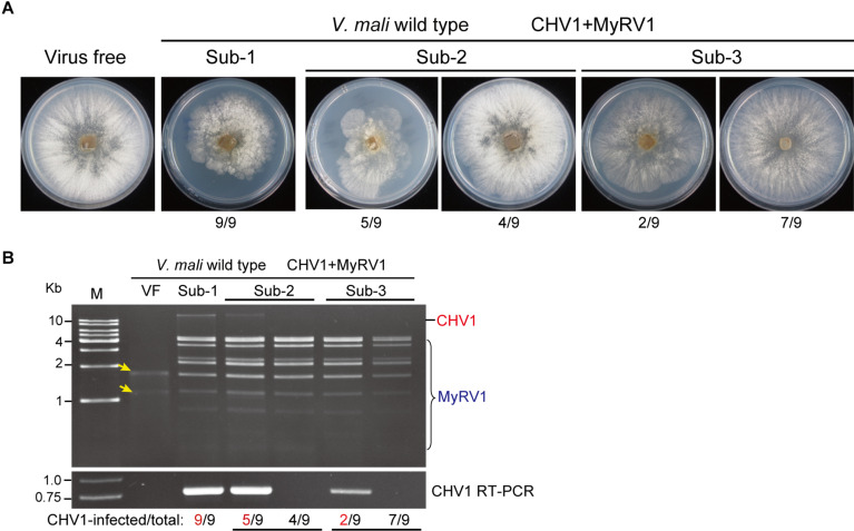
FIGURE 3.
The accumulation of CHV1 and MyRV1 in co-infected V. mali after subsequent subculturing. (A) The phenotypic growth on PDA medium of representative V. mali strains co-infected with CHV1 and MyRV1 in the first to third fungal subcultures (Sub-1 to Sub-3). The colonies were grown on PDA for 4 days and then photographed. The numbers below the images indicate the number of fungal colonies showing a similar phenotype as presented in the photo per total number of colonies shown. (B) Viral dsRNA and RT-PCR analyses of doubly infected V. mali strains described in (A). The numbers below the lanes indicate the number of samples in which CHV1 was detected or undetected per total number of samples. Yellow-colored arrows mark the traces of ribosomal RNAs in virus-free (VF) sample incorporated during dsRNA isolation.

